 | |||||||||||||||
 |
 |
 |
|
 |
|
 |
|
 |
|
 |
|
 |
|
 |
 |
V-CEM
VANDERBILT
CRYO
ELECTRON
MICROSCOPY
V-CEM Only:
Vanderbilt Cryo Electron Microscopy Facility
Overview
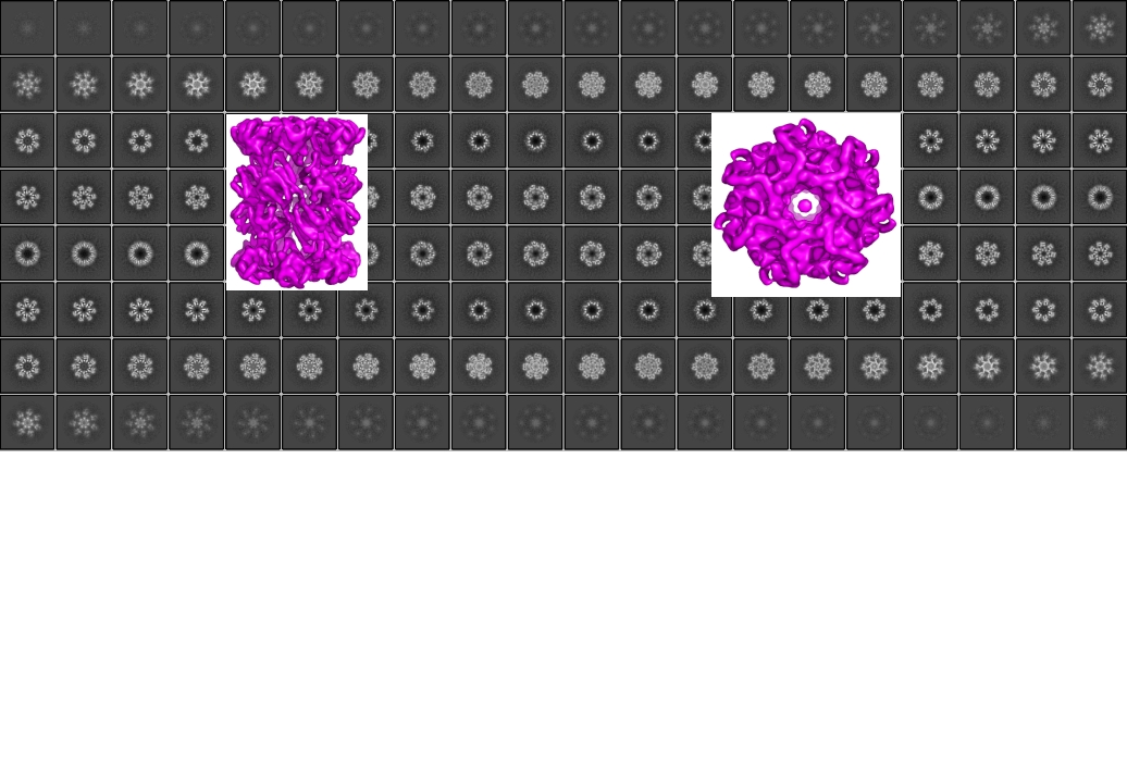
The V-CEM facility provides expertise and resources for the structural analysis of biological molecules using single particle analysis, tomography, and two-dimensional (2D) crystallization. The facility houses high-resolution transmission electron microscopes (TEMs) and cameras needed to collect data for single particle analysis and tomography.
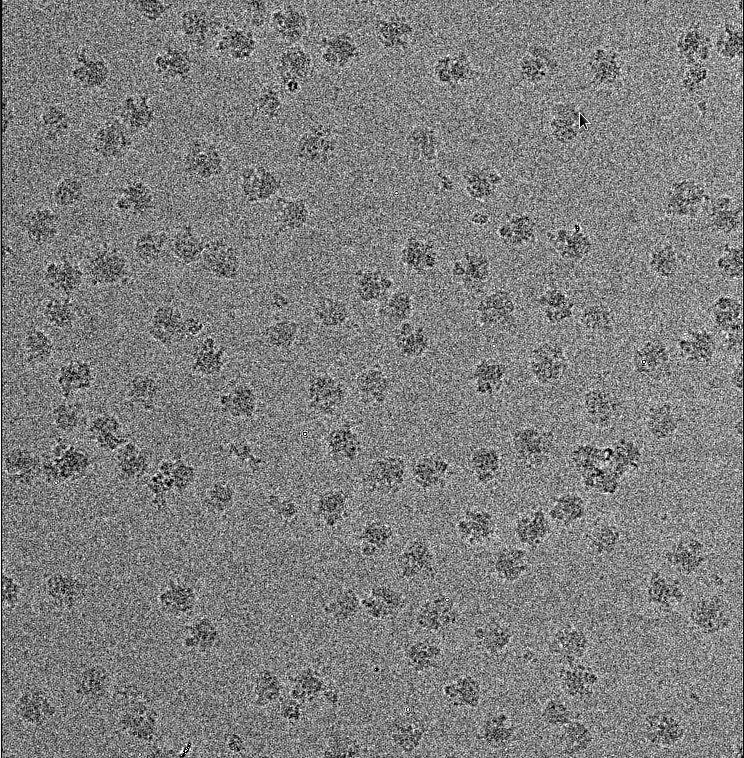
The facility houses a 300 kV FEI Polara equipped with a Gatan K2 Summit Direct Electron Detector (DED) camera, a 200 kV FEI F20 equipped with a Gatan Ultrascan 4K x 4K CCD, two FEI vitrobots (mark II\III), and the accompanying instrumentation needed for both negative stain and cryo-EM analysis. Biological samples are prepared for Molecular EM using either negative stain or vitrification (i.e. cryo-EM). For negative stain EM, the sample is embedded in a layer of dried heavy metal solution. For cryo-EM, the sample is suspended across a holey carbon grid and then rapidly plunged into cryogen, trapping the molecules in a thin layer of vitrified ice composed of the purification buffer. Negative stain provides high-contrast structural information mainly of the outer envelope of a molecule, while cryo-EM allows for the structural determination of near-atomic to atomic resolution 3D structures.

Mission Statement
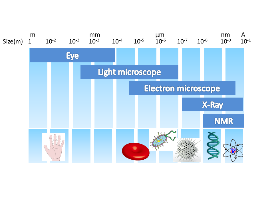
The goal of V-CEM is to make molecular EM accessible to all interested researchers at Vanderbilt. Molecular-EM is one of the fastest developing, cutting-edge structural techniques for exploring the organization of biological macromolecules. The unique strength of molecular EM is its versatility. Structures determined using molecular EM can span resolution ranges from very low- to atomic-levels (>30–1.8Å) and do not require crystalline samples for analysis.
In pursuit of its goal, the V-CEM enables molecular EM training and outreach by
- 1. Providing extensive training to new users.
- 2. Providing advice and support ranging from specimen preparation, data collection and image processing, computational image analysis, and structure determination.
|
VU Home |
VUMC Home |
People Finder |
University Calendar
webmaster- modified on September 04, 2019 |