
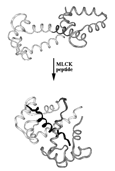
Change in quarternary structure upon binding of the myosin light chain kinase peptide to calcium-loaded calmodulin. Calmodulin N-terminal domain (residues 1-75_ is colored light grey, calmodulin C-terminal domain (residues 84-148) is colored medium grey, the linker between the two domains is colored dark grey, and the MLCK peptide is colored black. The molecular structures were rendered in Insight using PDB coordinates 1CLL (calcium-loaded calmodulin) and 2BBM (CaM-MLCK peptide complex)
This figure appeared as figure 10 in the book chapter:
Nelson, M.R., Chazin W.J., "Calmodulin as a calcium sensor" in
Calmodulin and Signal Transduction (1998) L.J. Van Eldik and
D.M. Watterson, eds. Academic Press, San Diego, pp. 17-64.
A color version is also available.
Details about file locations and color scheme. Internal access only
 |
 |
 |
 |
 |
 |
 |
 |
 |
CaBP Data Library Home Page|
General Info|
Sequence Info|
Structural Info|
Analysis Tools
Top of Current Section|
Search the Data Library|
Site Map|
Feedback