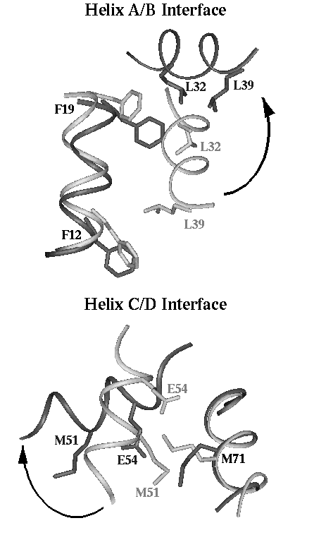
Opening of the EF-hands upon calcium binding to calmodulin. Illustration of the calcium-induced opening determined from the distance difference matrix and inter-residue contact analysis of the Helix A/B interface and Helix C/D interface in the N-terminal domain of calmodulin. The apo structure (shown in light grey) is from PDB coordinates 1CFC (model #1). The calcium-loaded structure (dark grey) is from PDB coordinates 1CLL. The apo and calcium-loaded structures were superimposed on the relatively stable A/D interface and rendered using Insight (MSI, San Diego). Examples of contacts made in only one of the two states are highlighted. In the A/B interface, Phe19 and L32, which are near the calcium-binding loop, contact only in the calcium-loaded state, while Phe12 and Leu39, found at the other end of the helices, contact only in the apo state. In the C/d interface, Met71 is different in the two states, contacting Glu54 only in the calcium-loaded state, and Met51 only in the apo state.
This figure appeared as figure 8 in the book chapter:
Nelson, M.R., Chazin W.J., "Calmodulin as a calcium sensor" in
Calmodulin and Signal Transduction (1998) L.J. Van Eldik and
D.M. Watterson, eds. Academic Press, San Diego, pp. 17-64.
There is also a similar color picture.
Details about file locations and color scheme. Internal access only
 |
 |
 |
 |
 |
 |
 |
 |
 |
CaBP Data Library Home Page|
General Info|
Sequence Info|
Structural Info|
Analysis Tools
Top of Current Section|
Search the Data Library|
Site Map|
Feedback