
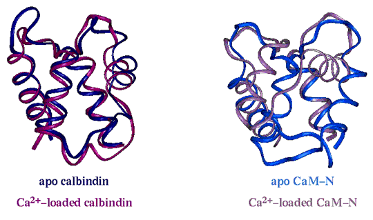
Structural response to calcium in calbindin D9k and calmodulin. Apo calbindin D9k is shown in dark blue. Calcium-loaded calbindin D9k is shown in red. Apo N-terminal domain of calmodulin is shown in light blue. Calcium-loaded N-terminal domain of calmodulin is shown in pink. As the picture indicates, the calcium-induced conformational changes are much more pronounced in the calmodulin domains than in calbindin D9k. The conformational changes in calmodulin involve rearrangements of the packing of the four helices in the domain. The conformational changes in calbindin D9k are much more subtle.
The molecular structures were rendered in Insight (MSI, San Diego) using PDB coordinates 1CLB (apo calbindin), 2BCB (calcium-loaded calbindin), 1CFC (apo calmodulin), and 1CLL (calcium-loaded calmodulin).
This figure was used in a poster presented at the 1997 International Symposium on Calcium-Binding Proteins and Calcium Function in Health and Disease.
Details about file locations and color scheme. Internal access only
 |
 |
 |
 |
 |
 |
 |
 |
 |
CaBP Data Library Home Page|
General Info|
Sequence Info|
Structural Info|
Analysis Tools
Top of Current Section|
Search the Data Library|
Site Map|
Feedback